The Art of Grafting: How Regenerative Solutions Facilitate Surgical Procedures
Preparation is key and seeing three steps ahead can be a game-changer in dental procedures. Before any dental implant placement takes place, dentists follow religiously a sequence of preparative actions – such as dental bone grafting. Used when the bone loss occurred in the jaw, a dental bone graft has the purpose of adding volume and density to the jaw area where the bone loss occurred. Providing the ideal background for the bone tissue to grow and regenerate, let’s dive into the particularities of this procedure.
A dental bone graft is exactly what it sounds like
It’s rather simple to define dental bone grafting: by making an incision in the jaw, a dentist/oral surgeon attaches other bone material to it, therefore providing additional support and preventing long-term oral health problems.
It is used as a filler and support, to enable the formation of new bone by acting as a mineral reservoir that induces its formation and at the same time to promote wound healing. These transplants are bioresorptive and do not have an antigen-antibody reaction.
Bone augmentation is possible due to the potential of bone tissue to fully regenerate if it is given the space in which it will grow. As bone tissue grows, it replaces biomaterial and fully integrates with the surrounding bone. After e.g. surgery, infection, tooth extraction due to various causes, trauma or advanced periodontal disease, the alveolar ridge changes in vertical and horizontal dimensions.
Three different processes are involved in bone regeneration – osteoconduction, osteogenesis and osteoinduction.
Osteoconduction is a process of bone replacement, ie. the granules serve only as a carrier for the disposal of newly formed bone (1, 2). Osteoblasts from the edge of the defect use the inserted material as a model to expand and create new bone. Basically, any material for bone regeneration should be at least osteoconductive. (3)
Osteogenesis involves the process by which vital osteoblasts from grafts contribute to new bone formation along with the following two described processes. (2, 3)
Osteoinduction is the process by which a graft encourages osteoprogenitor cells to differentiate into osteoblasts (2). The most common type of osteoinductive cell mediator is bone morphogenetic protein (BMP). Inserted material that is osteoconductive and osteoinductive not only serves as a model and support for currently existing osteoblasts, but encourages the creation of new osteoblasts, theoretically encouraging faster integration of the replacement itself. (4)
The term osteopromotion is often mentioned in the dental arena. It involves improving osteoinduction without possessing osteoinductive properties alone. For example, an enamel matrix derivative enhances the osteoinductive effect of demineralized lyophilized bone allograft (DFDBA), but will not stimulate bone growth alone. (5)
A bone graft can be made from your own body (autogenous), but it could also be obtained from a human tissue bank (allograft) or from an animal tissue bank (xenograft). Bone graft materials can in some cases be synthetic (alloplast).
Alloplastic materials can be produced from hydroxylapatite, a natural mineral that is also the main mineral component of bone. They can also be made of bioactive glass or calcium phosphate and are biologically active depending on the solubility in the environment. This type of material in combination with growth factors or bone marrow increases biological activity. Hydroxylapatite is a synthetic bone graft, which is mostly used today due to its osteoconductive and mechanical properties and biocompatibility. Today, tricalcium phosphate is also widely used in combination with hydroxylapatite to improve the effects of osteoconductivity and resorption. The advantage of alloplastic material is that it does not contain animal or human tissues or derivatives and is practically unlimited. (6)
How do you know you need one?
The most common reasons for having a dental bone graft are tooth loss or gum disease. You may need to undergo this procedure if:
- You are having a tooth extracted.
- Maxillary sinus elevation
- The missing tooth will be replaced by a dental implant (alveolar ridge preservation)
- Getting dentures requires the rebuilding of the jaw.
- Due to gum disease (periodontal disease), you have areas of bone loss.
Dental bone grafts: how does it work?
The bone graft material will be placed between the two sections of the bone that need to regenerate together. The next step is securing the bone graft with a regenerative solution and finally, the incision is sewn up to start healing.
In this procedure, regenerative solutions play a key element, being designed for the filling of bone voids or defects as well as enabling an optimal barrier effect.
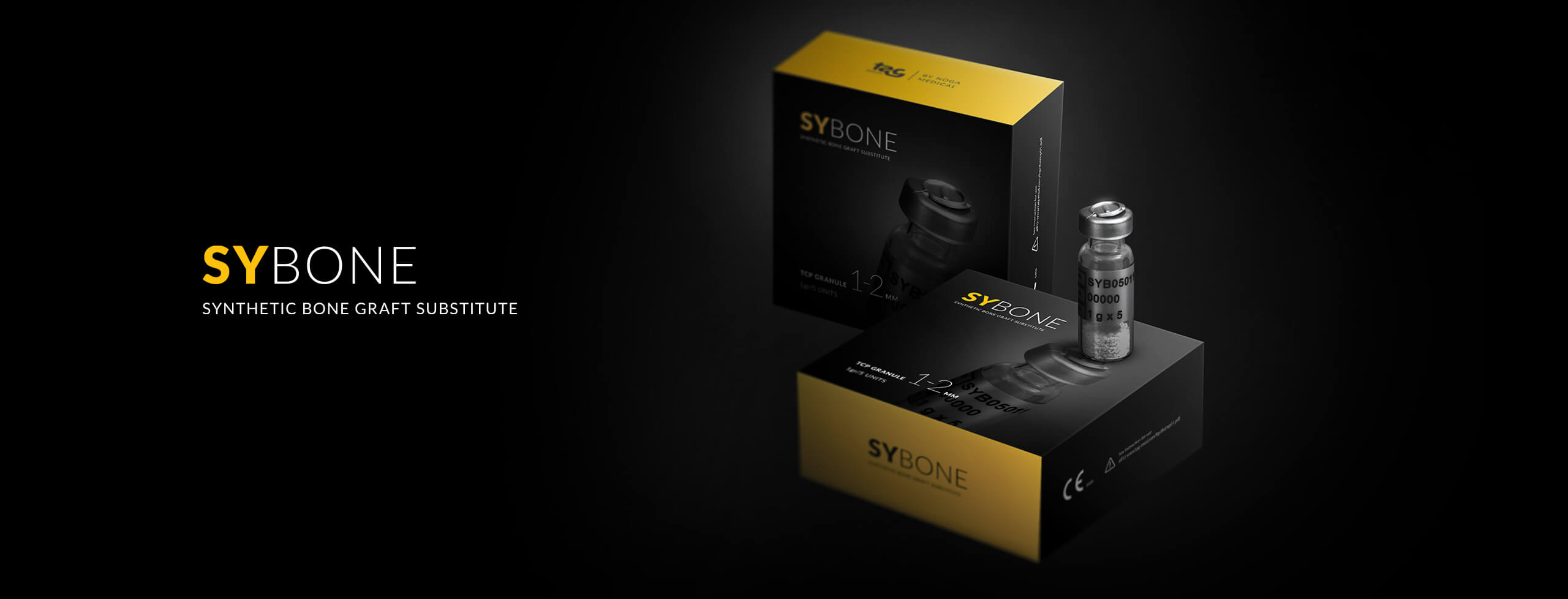
Thoughtfully crafted, TAG Dental’s line of regenerative solutions is designed to enable superior quality and consistent grafting. Our 100% synthetic porous osteoconductive ceramic, SyBone®TCP, provides excellent osteoconductive properties. The product contains 99% tricalcium phosphate, which fills bone defects with open porosity and provides high mechanical resistance. Among the benefits we count the zero-infection risk, great resorption time and availability in a variety of sizes, all while being made from 100% synthetic materials.
Angiogenesis is considered an important factor in the formation of bone structure. Due to the high porosity of SyBone®TCP biomaterial, better angiogenesis and the formation of harder, denser, and more vascularized bone are enabled. This condition allows the gradual resorption of alloplastic biomaterial, which in turn increases bone mass. As faster and greater new bone formation is encouraged, the use of a resorbable membrane was not necessary (a resorbable membrane would be indicated possibly in cases where there is possible material exposure).
Conclusion
The most common use of bone replacement in dentistry is during implant rehabilitation. Using popular materials (xenogeneic or alloplastic), we can achieve good angiogenesis and successful and predictable regeneration of bone mass at minimal cost, without a new operating field and creating additional discomfort for the patient.
To learn more about the latest innovation in implantology, find out more on our website or follow us on social media – Facebook, Instagram, Linkedin, Twitter.
We are looking forward to hearing your notes and thoughts about this topic.
Literature:
- Laurencin C, Khan Y, El-Amin SF. Bone graft substitutes. Expert Rev Med Devices. 2006 Jan; 3(1):49-57.
- Ehrler DM, Vaccaro AR. The use of allograft bone in lumbar spine surgery. Clin Orthop Relat Res. 2000 Feb;(371):38–45.
- Hung, Nguyen. (2012). Basic Knowledge of Bone Grafting. 10.5772/30442.
- Fee, L. Socket preservation. Br Dent J. 2017, 222, 579–582.
- Giannoudis PV, Dinopoulos H, Tsiridis E Injury. Bone substitutes: an update. 2005 Nov; 36 Suppl 3():S20-7.
- Zhao R, Yang R, Cooper PR, Khurshid Z, Shavandi A, Ratnayake J. Bone Grafts and Substitutes in Dentistry: A Review of Current Trends and Developments. Molecules. 2021 May 18;26(10):3007. doi: 10.3390/molecules26103007. PMID: 34070157; PMCID: PMC8158510.
